The human eye belongs to a general group of eyes found in nature called “simple eyes“. It contains a single lens that focuses light onto a light-sensitive membrane called the retina. Light-sensitive cells on retina convert light into electro-chemical signals, which are sent to brain via optic neurons to form the images that we see.
Structure of the eye. The cornea is a transparent structure found in the very front of the eye that allows light to go into the eyes. Most of the bending of light actually takes place as it goes through the cornea and the aqueous fluid (called Aqueous humor) in the anterior chamber between the cornea and the iris. This bending is possible because of the curve of the cornea as well as the different refractive indexes of air and that of the cornea and the aqueous fluid. Air has a refractive index of about 1.00, and the aqueous humor behind the cornea has a refractive index of about 1.33.
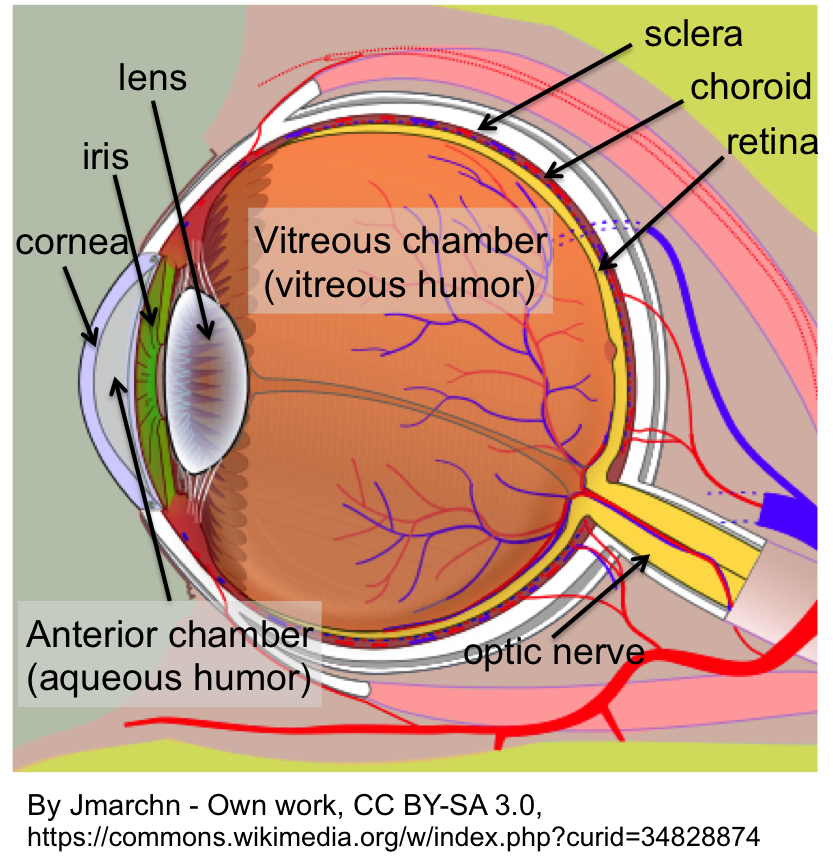
Behind the aqueous humor is a colored, ring-shaped membrane called the iris. It has an adjustable opening called the pupil (circular in human eyes, but may take different shapes in other animals), which can expand or contract to control the amount of light entering the eye. The pupil appears black because most light entering the eye through it gets absorbed and doesn’t come out. The iris contains two groups of smooth muscles; a circular group called the sphincter pupillae, and a radial group called the dilator pupillae. Contraction of the sphincter pupillae reduces the size of the pupil, while contraction of the dilator pupillae dilates the pupil.
Situated behind the pupil is a transparent structure called the crystalline lens. Ciliary muscles surround the lens and hold it in place. Furthermore, when the ciliary muscles relax, they pull on and flatten the lens, allowing the eyes to see objects that are far away. To see closer objects clearly, the ciliary muscle must contract in order to thicken the lens (this is why when our eyes get tired after reading a book for a long time). The interior vitreous chamber of the eyeball is filled with a jelly-like tissue called the vitreous humor. After passing through the lens, light must travel through this humor before striking the light-sensitive layer of cells called the retina, on which images are formed.
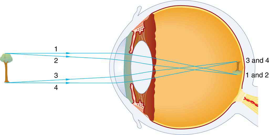
Taking an image with our eyes. The eyes work in exactly the same way as a camera. Light from an object gets focused by the cornea, aqueous humor, and lens onto the retina. By adjusting the shape of the lens, clear images of objects at different distances can be formed. Note that the images on retina are actually upside down, but our brain has evolved to adjust this so we see the world properly.
The retina is the innermost of three tissue layers that make up the eyeball. The outermost layer, called the sclera, is what gives most of the eyeball its white color. The middle layer between the retina and sclera is called the choroid. The choroid contains blood vessels that supply the retina with nutrients and oxygen and remove its waste products.
Embedded in the retina are millions of light sensitive cells, which come in two main varieties: rods and cones. Rods are used for monochrome vision in poor light, while cones are used for color and for the detection of fine detail. Cones are packed into a part of the retina directly behind the retina called the fovea, which is responsible for sharp central vision. When light strikes either the rods or the cones of the retina, it’s converted into an electric signal that is relayed to the brain via the optic nerve. The brain then translates the electric signals into the images that we see.
Watch this Youtube video for more details on how our eyes work.
Vision problems/diseases. The most common problems with vision are nearsightedness (myopia), farsightedness (hyperopia, caused by an irregularly-shaped eye, e.g. shorter eyeball, prevents light from properly lining up with the retina. The result is that it’s hard to see things close up. People of any age, including babies, can be farsighted.), astigmatism (a defect in the eye caused by nonspherical curvature) and age-related farsightedness (presbyopia, the lens of the eye gradually loses its flexibility, making it harder to focus on objects up close).